ARCHIVED - Cervical Cancer Screening in Canada: 1998 Surveillance Report
1. Introduction
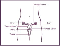
The cervix is the lower portion of the uterus leading into the vagina (Figure 1). It is lined with two main types of cell: squamous and glandular mucus-secreting cells. The junction between the two types of cell is called the transformation zone (or squamo-columnar junction) and is an area of rapid cell turnover where benign and malignant cellular changes are most likely to occur.
Cervical cancer is a malignancy of the cells lining the surface of the cervix. It begins as asymptomatic pre-cancerous lesions and usually develops gradually over many years. The intraepithelial lesions are limited to the cervical epithelium, and as invasion occurs the neoplastic cells penetrate the underlying membrane with potential for widespread dissemination. Depending on their severity, lesions can resolve on their own or can progress to cancer. Cervical cancers most commonly arise from the squamous cells (70%), and 18% to 20% arise from the glandular cells (adenocarcinomas). Adenosquamous carcinomas (5%) share features of squamous cell carcinomas as well as adenocarcinomas, but rarely occur. Other unspecified type of cervical cancer account for the remaining 5%Footnote 1.
Figure 1: Female Reproductive System

Cervical cancer affects women of all age ranges, but the highest incidence is found among women aged 40-59Footnote 1. Women with cervical cancer show a relatively good prognosis: the ratio of deaths to cases is 0.29, and the 5-year relative survival rate is 74%Footnote 2. Effective treatments for cervical cancer are readily available.
Because of the natural history of cervical cancer, specifically the presence of pre-cancerous stages that can be easily detected and treated, the disease lends itself well to screening programs. The Papanicolaou (Pap) test is an established method used to examine cells obtained from the cervix in order to determine whether they show any signs of pre-cancerous changes. A spatula and/or brush is used to sample cells from the transformation zone (squamo-columnar junction), which are then smeared onto a glass slide and examined under the microscope by a cytotechnologist for any type of pre-cancerous changes. The cytopathologists and cytotechnologists classify the cells according to a spectrum from normal to carcinoma.
Several different classification schemes have evolved over the years for characterizing Pap test results. The CIN (cervical intraepithelial neoplasia) grading system has been gradually replaced by the Bethesda System (introduced in 1989)Footnote 3, although some provinces in Canada continue to use the CIN grading system. Appendix B provides a comparison between the various nomenclature used to classify cell abnormalities seen on Pap tests.
In Canada, opportunistic screening has occurred since the introduction of the Pap test and is by far the most frequent way in which women receive screening services. Pap smears are taken by general practitioners, gynecological specialists and, in some circumstances, nurses in doctors' offices, community health clinics and hospitals.